
1. Introduction
Hyperuricemia and hypertension are interlinked conditions that significantly impact cardiovascular and renal health [1]. Hyperuricemia is clinically defined as serum uric acid (SUA) levels exceeding 7.0 mg/dl in men and 6.0 mg/dl in women, resulting from either increased uric acid production or decreased renal excretion. Hypertension, defined as blood pressure of 140/90 mm Hg or higher, remains a major contributor to global morbidity and mortality. Early epidemiological studies established a strong association between elevated SUA levels and hypertension [2].
Recent meta-analyses indicate that hyperuricemia is an independent risk factor for incident hypertension, particularly in younger demographics and women [3]. A 1 mg/dl increase in SUA elevates the risk of developing hypertension by approximately 13%, independent of age, body mass index (BMI), renal function, or diabetes status [4]. Multiple studies corroborate the role of SUA as a reliable predictor of future blood pressure elevation [5].
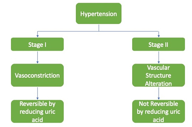
Current evidence positions hyperuricemia as a modifiable risk factor in the management of hypertension [6]. Uric acid–lowering therapies, such as allopurinol, have demonstrated efficacy in improving blood pressure control among newly diagnosed hypertensive patients, notably in adolescents and young adults [7]. The pathogenic effects of uric acid occur in two distinct phases [8]. The initial phase is reversible, involving reduced nitric oxide (NO) bioavailability and renin-angiotensin system (RAS) activation, which impairs vascular compliance [9]. The secondary phase involves irreversible vascular changes, including smooth muscle hypertrophy and structural remodeling, sustaining hypertension even subsequent to SUA normalization [10].
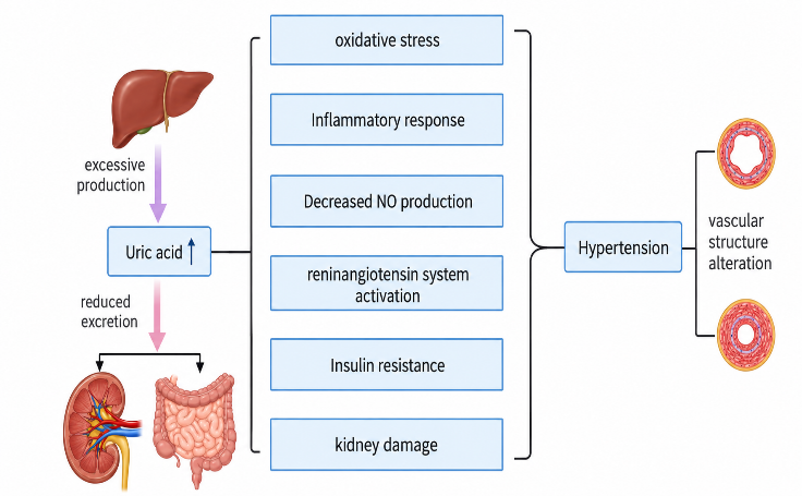
Multiple concurrent mechanisms—oxidative stress, inflammation, endothelial dysfunction, renal microvascular damage, RAS activation, insulin resistance, and vascular structural changes—synergistically produce hyperuricemia-related hypertension [11]. Comprehending these interconnected pathways is critical for developing targeted preventive and therapeutic interventions [12].
The increasing prevalence of hypertension in adolescents and young adults is a recognized global health concern. While clinical focus often centers on cardiovascular diseases, neurological disorders such as stroke necessitate attention due to long-term socioeconomic impacts. Strokes in younger adults account for 10-15% of total cases but pose significant challenges as they occur during prime working years. Unique risk factors—hypertension, diabetes, dyslipidemia, smoking, and obesity—demand age-tailored interventions. Elevated serum uric acid has emerged as an early indicator for the prevention of cardiovascular and cerebrovascular diseases [13]. Furthermore, hypertension within the portal venous system represents a significant liver disorder linked to elevated vascular resistance caused by fibrosis and chronic liver injury, leading to complications like ascites and hepatic encephalopathy. Timely diagnosis is vital for preventing systemic complications [14].
2. Uric Acid and Hypertension
Hyperuricemia denotes excessive levels of uric acid in the bloodstream [15]. This condition is intimately linked to the pathogenesis of primary hypertension, often presenting prior to the clinical onset of elevated blood pressure [16]. Both conditions are exacerbated by modern dietary habits, metabolic disorders, and sedentary lifestyles [17]. High fructose consumption plays a crucial role by boosting uric acid production, alongside driving weight gain, insulin resistance, and hyperlipidemia [18]. The correlation between uric acid and blood pressure is most robust in younger populations; as age advances, structural changes in blood vessels and kidneys alter the dynamic of this connection [19]. Notably, infants born with low birth weight often possess fewer nephrons, increasing their susceptibility to essential hypertension later in life [20].
2.1 Mechanisms of Hyperuricemia-Induced Hypertension
Elevated uric acid serves as an early biomarker for renal impairment and microalbuminuria [21]. Clinicians increasingly utilize it as an early alert and a therapeutic target for blood pressure management [22]. Pharmacological agents like allopurinol effectively lower uric acid and assist in blood pressure regulation, demonstrating particular efficacy in adolescents with nascent hypertension [23]. At the cellular level, uric acid triggers hypertension via interconnected pathophysiological pathways [24], encompassing oxidative stress, vascular smooth muscle proliferation, endothelial dysfunction, inflammation, and RAS activation [25]. These cascades induce vascular stiffness and escalate peripheral flow resistance [26]. The primary mechanisms are elucidated below [27].
2.1.1 Oxidative Stress
Oxidative stress occurs when the generation of reactive oxygen species (ROS) overwhelms intrinsic antioxidant defenses [28]. These deleterious molecules damage essential cellular macromolecules, including lipids, proteins, and DNA, resulting in cellular instability [29]. Chronic oxidative imbalance is a central feature of cardiovascular and metabolic diseases [30]. Uric acid exhibits a paradoxical role in redox homeostasis: acting as a protective antioxidant in the extracellular fluid under normal physiological conditions, but transforming into a potent pro-oxidant when it accumulates intracellularly [31][33].
Excessive intracellular uric acid inhibits mitochondrial energy metabolism, stimulates ROS production, and disrupts critical signaling cascades [34]. While physiological levels of extracellular uric acid preserve endothelial integrity by preventing oxidation and facilitating NO-mediated vasodilation [35], supraphysiological concentrations induce endothelial dysfunction characterized by diminished NO, arterial stiffening, and impaired vasorelaxation [36].
In hyperuricemic states, the metabolic generation of uric acid via xanthine oxidase simultaneously produces substantial quantities of ROS [37][38]. Furthermore, uric acid activates NADPH oxidase, amplifying superoxide production and exacerbating cellular injury [39]. Uric acid also induces mitochondrial dysfunction, triggering mitochondrial ROS generation, depleting ATP, and compromising cellular bioenergetics [40]. Intracellularly, uric acid activates the MAPK signaling pathway (specifically p38 MAPK), which further upregulates NADPH oxidase, propagates inflammation, and accelerates vascular remodeling [41].
Clinical and experimental data firmly link elevated serum uric acid with systemic hypertension and vascular pathology [42]. Elevated levels impair NO bioavailability, compromise vasodilation, and stimulate the proliferation of vascular smooth muscle cells (VSMCs), increasing systemic vascular resistance [43]. Xanthine oxidase inhibitors, such as allopurinol and febuxostat, mitigate this oxidative burden and lower systemic and renal blood pressure [44]. The concomitant generation of ROS from NADPH oxidase and dysfunctional mitochondria accelerates apoptosis, cellular senescence, and atherogenesis, perpetuating a vicious cycle of vascular injury [45][49].
2.1.2 Inflammatory Activation
Elevated uric acid (UA) levels are strongly correlated with systemic inflammation, evidenced by positive associations with inflammatory biomarkers such as C-reactive protein (CRP) and interleukin-6 (IL-6). UA propagates inflammation via three primary intracellular signaling pathways:
-
MAPK Pathway: UA activates the mitogen-activated protein kinase (MAPK) pathway. It stimulates p38 MAPK and p44/42 MAPK in a time- and dose-dependent manner, promoting the release of inflammatory cytokines. UA also triggers extracellular signal-regulated kinase (ERK), supporting the secretion of chemokines and cell adhesion molecules.
-
AMPK Suppression: UA suppresses the AMP-activated protein kinase (AMPK) pathway. Since AMPK typically inhibits the NLRP3 inflammasome, its suppression by UA facilitates NLRP3 activation and heightens the inflammatory response.
-
PI3K/Akt Pathway: UA activates the PI3K/Akt pathway, upregulating mTOR activity, reducing autophagy, and enhancing inflammation and angiogenesis via hypoxia-inducible factor-1α (HIF-1α) upregulation.
In endothelial cells, UA induces the expression of CRP, IL-6, IL-8, Angiotensin II (Ang II), TNF-α, ICAM-1, and VCAM-1. In VSMCs, UA promotes proliferation and migration. Elevated chemokines encourage monocyte adhesion and infiltration into the subendothelial space, causing endothelial dysfunction. When serum UA exceeds ~7.0 mg/dL, sodium urate crystals may precipitate, activating macrophages and neutrophils via TLR2/4 and CD14. This triggers MyD88/TRIF signaling, NF-κB activation, NLRP3 inflammasome assembly, and the release of IL-1β [50].
These inflammatory mechanisms directly contribute to hypertension by increasing vascular permeability, inducing cytokine-driven vasoconstriction, stiffening resistance vessels, and stimulating sodium and water retention.
2.1.3 Reduced Nitric Oxide (NO) Bioavailability
Nitric oxide (NO) is critical for endothelial homeostasis, acting as a potent vasodilator and antioxidant. NO regulates vascular tone, supports angiogenesis, inhibits leukocyte adhesion, and prevents platelet aggregation. Hyperuricemia diminishes NO bioavailability through three primary mechanisms: exacerbating oxidative stress, depleting L-arginine supplies, and directly inhibiting endothelial nitric oxide synthase (eNOS).
Elevated uric acid directly impacts the macula densa of the kidney, lowering NO synthase levels and inducing endoplasmic reticulum stress via the protein kinase C/eNOS pathway. This diminishes eNOS activity, leading to VSMC constriction. The reduction in NO consequently upregulates angiotensinogen, ACE, and Ang II receptors, thereby overactivating the RAS and causing pathological vasoconstriction. Furthermore, hyperuricemia upregulates arginase in endothelial cells, diverting L-arginine into the urea cycle and away from NO synthesis. Research demonstrates that high uric acid prevents eNOS from coupling with calmodulin, further attenuating NO production [51].
2.1.4 Renin–Angiotensin System (RAS) Activation
Uric acid actively stimulates the RAS, particularly the ACE-Ang II–AT1 receptor axis, leading to vasoconstriction and hypertension. Experimental models demonstrate that RAS inhibitors (e.g., enalapril, losartan) effectively avert hyperuricemia-induced hypertension and arteriolar damage. In VSMCs, UA promotes proliferation by stimulating local RAS components via increased expression of platelet-derived growth factor (PDGF) and thromboxane synthesis, activating specific MAPK pathways. UA synergizes with Ang II to amplify vascular inflammation, oxidative stress, and structural remodeling [52].
2.1.5 Insulin Resistance
Significant epidemiological evidence links elevated UA levels with insulin resistance (IR). UA impairs insulin-mediated eNOS activation, disrupting the PI3K/Akt signaling pathway and dampening NO output. Peripheral tissue IR plays a dominant role in hyperuricemic hypertension. UA and IR interact synergistically to elevate blood pressure by:
-
Hyperactivating the RAS
-
Increasing renal sodium reabsorption
-
Impairing endothelial NO regulation [53]
Urate-lowering therapies, such as xanthine oxidase inhibitors, have been shown to ameliorate IR and systemic inflammation, validating the mechanistic linkage.
2.1.6 Renal Injury
Animal models demonstrate that hyperuricemia induces hypertension via primary damage to preglomerular arterioles. Chronic elevation of uric acid induces renal oxidative stress, mitochondrial damage, and ATP depletion. This disrupts renal autoregulation of blood flow. The resultant renal ischemia triggers inflammatory and fibrotic cascades within the renal tubulointerstitium, driving blood pressure higher. UA also stimulates endothelin-1 production and fibroblast proliferation, perpetuating renal microvascular disease and establishing a feedback loop that sustains hypertension [54][55].
3. Pathophysiology
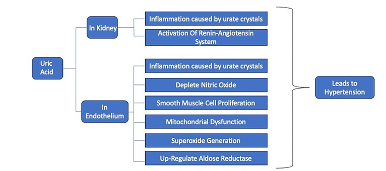
Hyperuricemia induces hypertension through complex mechanisms involving renal microvascular damage and endothelial dysfunction.
3.1.1 Renal Impact
Elevated uric acid damages renal architecture. Urate precipitation and localized RAS activation in the kidneys cause cellular inflammation, tubular injury, and fibrosis. RAS activation promotes sodium and water retention, raising blood pressure. Subsequent chronic renal architectural damage ensures that hypertension persists even if uric acid levels are therapeutically normalized [56].
3.1.2 Endothelial Impact
Uric acid impairs vascular function through:
-
Oxidative stress: Generating ROS that quench NO, inhibiting vasodilation.
-
Intracellular toxicity: Entering endothelial cells via specific transporters, impairing mitochondrial ATP synthesis, and upregulating oxidative enzymes, leading to vascular stiffness [57].
-
Polyol pathway activation: Soluble uric acid stimulates aldose reductase, further diminishing NO and driving endogenous fructose production, exacerbating endothelial dysfunction [57].
3.1.3 Phases of Hyperuricemia-Induced Hypertension
The progression of hyperuricemia-induced hypertension occurs in two distinct clinical stages:
-
Uric acid-dependent stage: Characterized by reversible vasoconstriction due to blocked NO and active RAS. Urate-lowering treatments can successfully normalize blood pressure.
-
Uric acid-independent stage: Characterized by permanent vascular structural alteration, including VSMC proliferation and arteriolosclerosis. This causes persistent hypertension that does not resolve solely with uric acid reduction [56][58].
4. Hyperuricemia, Inflammation, and Hypertension
4.1 Nonimmune Mechanisms (First Hit)
In the initial stage, uric acid exerts direct, immune-independent damage on renal and vascular tissues [59][60]. This involves activation of the RAAS and inhibition of eNOS, leading to systemic vasoconstriction and sodium retention [61]. Intracellular UA accumulation activates NADPH oxidase, causing mitochondrial dysfunction and oxidative stress [62], which drives VSMC proliferation [63] and increases sympathetic nervous system tone, raising overall peripheral resistance [64].
4.2 Immune Mechanisms (Second Hit)
In the chronic phase, the disease transitions to an immune-driven state [65]. Uric acid acts as a Damage-Associated Molecular Pattern (DAMP), activating macrophages and dendritic cells [66]. Intracellular uptake of UA triggers the NLRP3 inflammasome, releasing potent inflammatory cytokines (IL-1β, IL-18) [67]. This results in sterile vascular inflammation and irreversible arterial remodeling [68]. Additionally, UA binds to Toll-like receptors (TLRs), triggering NF-κB signaling [69]. This immune activation constitutes the "second hit," perpetuating vascular stiffness and organ damage regardless of acute uric acid levels [70][71].
5. Future Perspectives
Future research must prioritize identifying the precise cardiovascular threshold of serum uric acid to clarify when UA begins exerting pathogenic vascular effects [72]. Current hyperuricemia criteria are based on urate solubility (relevant to gout) rather than cardiovascular risk [73][74]. Understanding the longitudinal fluctuation of UA is critical, as transient spikes may promote cumulative endothelial injury [75][77]. Furthermore, the development of advanced biomarkers—such as urinary urate excretion and xanthine oxidase activity—is required, as standard serum UA fails to capture intracellular metabolic stress [78][79]. Integrating these approaches will guide early intervention strategies to mitigate cardiovascular risk [80][81].
6. Conclusion
Hyperuricemia is increasingly recognized as a profound metabolic and cardiovascular risk factor extending far beyond gout. Elevated serum uric acid is mechanistically linked to primary hypertension, coronary artery disease, and cerebrovascular pathology. Uric acid's dual role—acting as a physiological antioxidant extracellularly but a pathological pro-oxidant intracellularly—drives endothelial dysfunction, oxidative stress, and systemic inflammation. The robust epidemiological and mechanistic link between uric acid and blood pressure underscores the utility of early clinical intervention, including dietary modification (reduced fructose/purines) and pharmacological management (xanthine oxidase inhibitors). Regular monitoring of serum uric acid should be integrated into comprehensive cardiovascular risk assessments to effectively reduce disease burden.
7. Recommendations
-
Routine Clinical Screening: Integrate serum uric acid measurements into standard hypertension assessments, particularly for patients with metabolic syndrome, obesity, or early-stage hypertension.
-
Risk Stratification: Utilize elevated UA levels as an early biomarker to identify high-risk patients requiring closer monitoring and aggressive lifestyle intervention.
-
Comprehensive Diagnostics: Evaluate SUA in conjunction with standard cardiac and renal panels to formulate holistic, targeted treatment regimens.
-
Lifestyle Modifications: Counsel hyperuricemic and hypertensive patients on dietary changes, emphasizing adequate hydration, weight management, and significant reduction of dietary fructose and purines.
-
Pharmacological Management: Consider urate-lowering therapies (e.g., allopurinol) in cases of refractory hyperuricemia and nascent hypertension, guided by emerging clinical evidence.
8. Declarations
Ethics Approval
Not applicable
Funding
No specific funding was received from any funding agencies in the public, commercial, or not-for-profit sectors for the preparation of this review article.
Conflicts of Interest
The authors declare that they have no known competing financial interests or personal relationships that could have appeared to influence the work reported in this paper.
Consent to Participate
Not applicable
Authors' Contributions
All authors contributed to the study conception and design. Literature search and data analysis were performed by B. Harshitha, K. Sushma, and M. Vandhana. The first draft of the manuscript was written by Dr. G. Ramya and B. Harshitha, and all authors commented on previous versions of the manuscript. V. Umamaheshwar Rao provided critical revision and overall supervision. All authors read and approved the final manuscript.