
Introduction
Spontaneous coronary artery dissection (SCAD) is an increasingly recognized cause of acute coronary syndrome (ACS) and sudden cardiac death, disproportionately affecting young to middle-aged women lacking classic atherosclerotic risk factors [1][2]. The clinical spectrum of SCAD is highly variable, ranging from mild angina to unstable hemodynamics, transmural myocardial infarction, ventricular arrhythmias, and cardiogenic shock [3][4]. Pathophysiologically, SCAD arises from a non-iatrogenic, non-traumatic tearing of the coronary arterial wall, creating a false lumen that propagates along the vessel and compresses the true lumen, leading to myocardial ischemia [5].
Invasive coronary angiography (CAG) remains the standard initial diagnostic modality; however, its reliance on two-dimensional luminograms presents significant limitations. Angiographic features of SCAD can mimic coronary vasospasm, thromboembolism, or conventional atherothrombotic disease, frequently leading to misdiagnosis or delayed recognition [5][6]. Moreover, catheter manipulation and intra-arterial contrast injection carry a heightened risk of precipitating or extending iatrogenic coronary artery dissection in this fragile vascular cohort [7].
With recent advancements in spatial and temporal resolution, multi-detector coronary computed tomography angiography (CTA) has emerged as an invaluable noninvasive alternative [8][9]. CTA provides direct visualization of the arterial wall cross-section, allowing definitive identification of intramural hematoma (IMH), intimal flaps, and perivascular inflammatory changes [9]. This case report highlights the diagnostic utility of coronary CTA in establishing the initial diagnosis and confirming complete anatomical resolution during longitudinal follow-up in a patient with SCAD.
Case Report
A 38-year-old male with no known past medical history or traditional cardiovascular risk factors presented to the emergency department following the sudden onset of severe, crushing left-sided chest pain radiating to the interscapular region. The symptoms occurred at rest and were accompanied by transient dizziness and diaphoresis. Upon arrival, the patient was distressed. Vital signs demonstrated a blood pressure of 124/78 mmHg, a heart rate of 175 beats per minute, and an oxygen saturation of 98% on ambient air. Physical examination revealed clear lung fields and normal heart sounds without murmurs or gallops.
The initial electrocardiogram (ECG) documented a sustained monomorphic ventricular tachycardia, which was promptly terminated via synchronized direct current cardioversion (150 J), restoring normal sinus rhythm. A post-cardioversion 12-lead ECG revealed ST-segment elevation in leads II, III, and aVF with reciprocal ST-segment depression in leads I and aVL, alongside dominant R waves and ST-segment depression in V1–V3, indicative of an acute inferior-posterior ST-segment elevation myocardial infarction (STEMI). Emergency transthoracic echocardiography demonstrated regional wall motion abnormalities (akinesis of the inferior and posterior segments) with an estimated left ventricular ejection fraction of 40% and no significant valvular dysfunction.
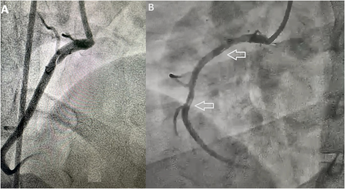
The patient was immediately transferred to the cardiac catheterization laboratory. Emergent CAG via a right radial approach demonstrated a long, linear radiolucent line extending from the proximal to the mid-segment of the right coronary artery (RCA), consistent with a Type 1 SCAD morphology. Antegrade coronary perfusion was maintained with TIMI 3 flow. The left anterior descending (LAD) and left circumflex (LCX) arteries were angiographically normal, free of luminal stenosis or atherosclerotic disease. Given the patient's hemodynamic stability, complete resolution of chest pain following medical stabilization, and preservation of distal vessel flow, a conservative, non-interventional management strategy was elected. Pharmacotherapy was initiated with dual antiplatelet therapy (aspirin 100 mg daily and clopidogrel 75 mg daily), metoprolol succinate, perindopril, and atorvastatin.
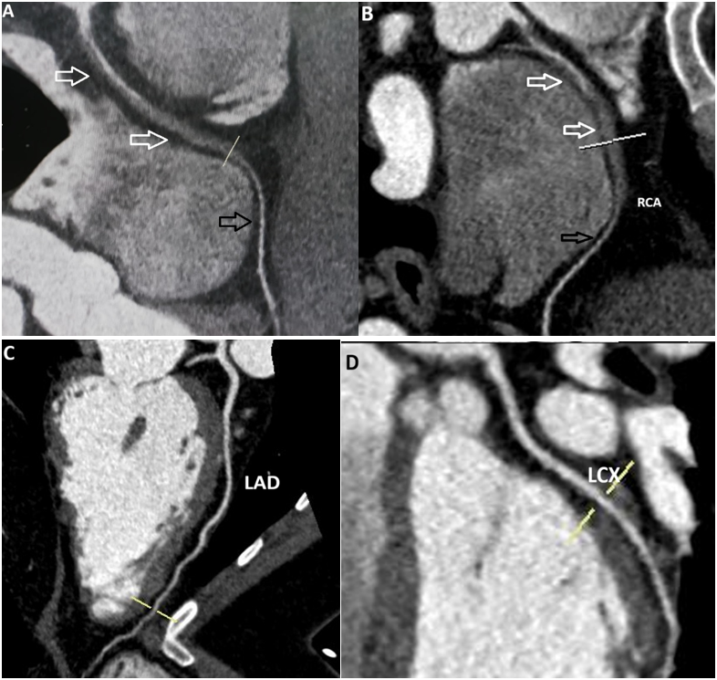
To establish a noninvasive baseline and further characterize the vessel wall, a 128-slice multi-detector coronary CTA was performed prior to discharge. The scan corroborated the angiographic findings, clearly demonstrating a non-calcified intimal flap and an extensive intramural hematoma causing eccentric luminal narrowing of the proximal-to-mid RCA, without extension into distal vessel branches. The patient had an uneventful clinical course and was discharged on day 5.
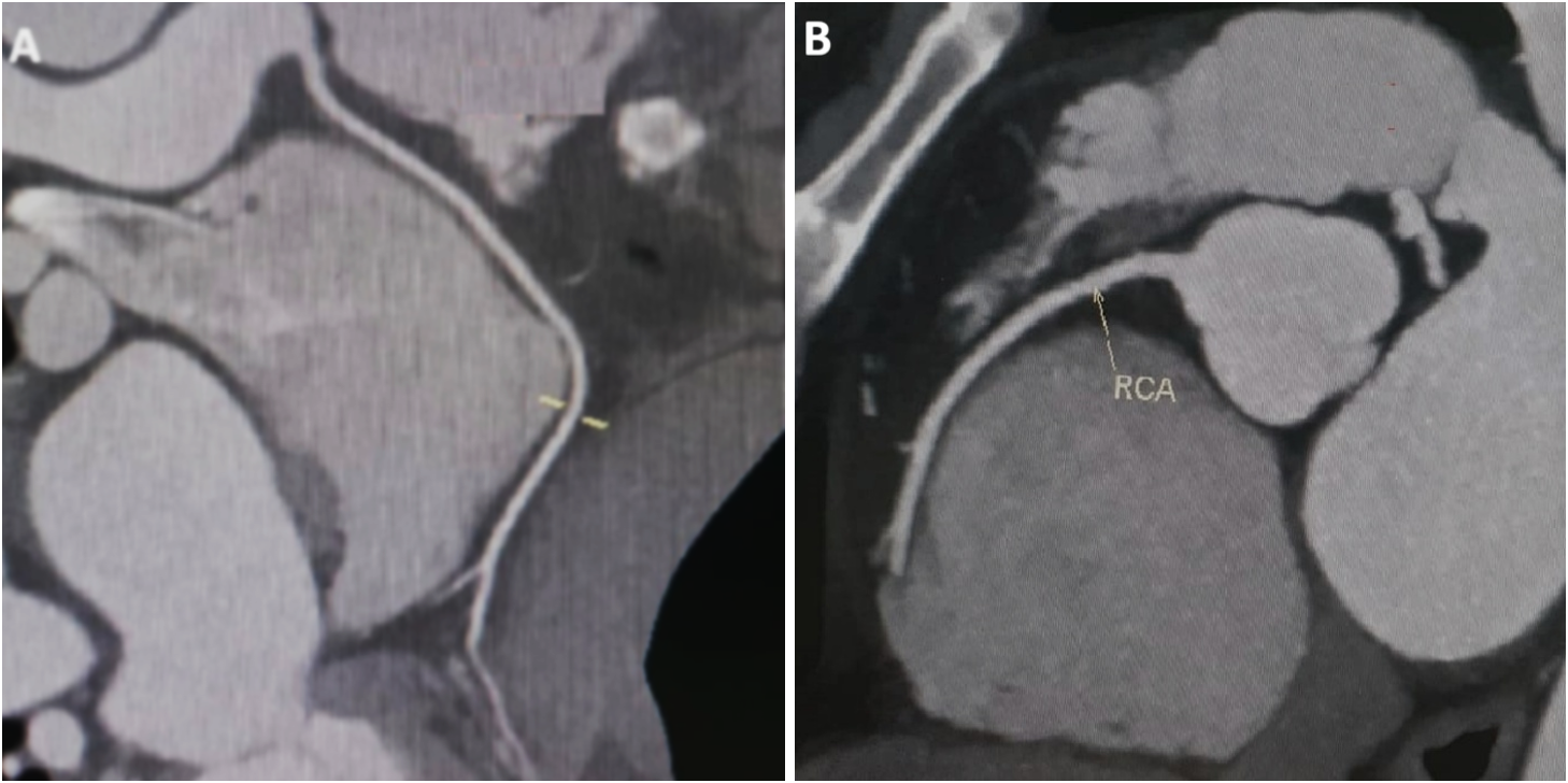
At the 12-month outpatient follow-up, the patient reported intermittent, atypical non-exertional chest discomfort. Physical examination and resting ECG were unremarkable. To assess vascular integrity without subjecting the patient to the risks of invasive catheterization, a repeat coronary CTA was performed. The follow-up scan revealed complete resolution of the intramural hematoma and intimal flap, with full restoration of normal RCA luminal caliber and architecture. The noninvasive imaging strategy confirmed complete spontaneous vascular healing, reassuring the patient and avoiding unnecessary repeat invasive angiography.
A baseline coronary (CTA) during hospitalization corroborated the dissection flap from proximal to mid/distal RCA segments, with normal distal RCA filling including the posterior descending artery and posterolateral branches. The patient was discharged in stable condition.
One-year follow-up coronary CTA, prompted by atypical chest pain, revealed complete resolution of the dissection with normal coronary anatomy. Noninvasive coronary CTA obviated repeat invasive angiography avoiding iatrogenic dissection risk, while confirming healing and establishing its pivotal role in SCAD diagnosis and surveillance.
Discussion
SCAD is an underdiagnosed cause of ACS, accounting for up to 4% of all ACS presentations overall and up to 35–50% of ACS cases in women under 55 years of age [1][7][8]. While overwhelmingly predominant in females, SCAD can occur in males, who often present following intense physical or emotional stressors [12][13].
Pathophysiology and Vascular Involvement
The underlying pathophysiology of SCAD involves either a primary intimal tear resulting in a secondary intramural hematoma, or primary rupture of vasa vasorum leading to hematoma formation within the tunica media [5][12]. This hematoma expands, creating a false lumen that compresses the true lumen and restricts myocardial blood flow. SCAD most commonly affects a single vessel (90–95% of cases), with the LAD being the most frequently implicated artery (40–70%), followed by the RCA (15-20%) and LCX (10–15%) [2][7][12]. Dissections predominantly affect mid-to-distal vessel segments; primary proximal involvement, as observed in our patient's RCA, is comparatively infrequent [7][13].
Diagnostic Modalities: CAG vs. Coronary CTA
Invasive coronary angiography remains the first-line investigation in acute presentation; however, it has key limitations. Saw et al. classified SCAD into three distinct angiographic phenotypes [11]:
-
Type 1 (25–30%): Classical radiolucent flap with contrast staining of the vascular wall.
-
Type 2 (50–60%): Smooth, diffuse luminal narrowing (>20 mm) caused by intramural hematoma without a visible intimal flap. Type 2a is flanked by normal proximal and distal segments, whereas Type 2b extends to the vessel terminus.
-
Type 3 (<5%): Focal or tubular stenosis (<20 mm) that mimics atherosclerotic stenosis, requiring intravascular imaging (IVUS or OCT) for confirmation.
Coronary CTA has emerged as a valuable complementary modality [14][15]. Modern multi-detector CTA platforms offer high spatial and temporal resolution, enabling direct visualization of cross-sectional vessel morphology [15][16]. Specific diagnostic features of SCAD on coronary CTA include [15][17]:
-
Non-calcified intramural hematoma presenting as eccentric wall thickening.
-
Presence of a distinct intimal flap separating true and false lumens.
-
Abrupt or tapered luminal attenuation without atherosclerotic plaque elsewhere.
-
Perivascular epicardial fat stranding, reflecting acute vascular inflammation.
Despite these advantages, coronary CTA possesses inherent technical limitations. Its diagnostic yield diminishes in distal vessel segments or side branches with diameters <2 mm due to motion artifacts and spatial resolution constraints [15][18]. Consequently, a negative CTA cannot completely exclude SCAD in small distal vessels.
Therapeutic Strategies and Follow-up Paradigms
Current consensus guidelines strongly support conservative medical management for hemodynamically stable patients with SCAD and preserved distal flow [20][21]. Observational studies show that spontaneous vascular healing occurs in over 70–90% of conservatively managed patients within months [4][7][20]. Revascularization via percutaneous coronary intervention (PCI) or coronary artery bypass grafting (CABG) is associated with high technical failure rates (up to 30-40%) and complication risks, including dissection propagation and stent malfunction [20][24][25]. PCI is strictly reserved for patients with ongoing ischemia, hemodynamic instability, or left main trunk involvement [21][22].
Given the risk of early recurrent events and the necessity of verifying vascular healing, longitudinal follow-up is critical [20][23]. Historically, repeat CAG was performed to evaluate persistent or recurrent symptoms. However, given the 2–5% risk of iatrogenic dissection extension during catheterization in SCAD patients [7], noninvasive coronary CTA represents a safer follow-up modality [15][16]. As demonstrated in this case, coronary CTA can definitively document complete resolution of the intramural hematoma and restoration of normal coronary anatomy, providing clinical reassurance without exposing the patient to procedural hazards [15].
Conclusion
Spontaneous coronary artery dissection is an important cause of acute coronary syndrome in patients without classic cardiovascular risk factors. While invasive coronary angiography remains essential in the acute diagnostic phase, coronary computed tomography angiography provides unique noninvasive advantages by directly visualizing intramural vessel wall pathology. Coronary CTA is a safe, highly effective modality for assessing stable patients with equivocal angiographic findings and for conducting long-term surveillance to confirm spontaneous vascular healing.
Declarations
Ethics Approval and Consent to Participate
Ethical approval for this case report was obtained from the Prince Sultan Cardiac Center-Qassim.
Consent for Publication
Written informed consent was obtained from the patient for the publication of this case report and any accompanying clinical images.
Availability of Data
Data are not publicly available to protect patient confidentiality but are available from the corresponding author on reasonable request.
Competing Interests
None
Funding
None
Authors' Contributions
All authors have equal contribution.
Acknowledgements
None