
1. Introduction
Uterine fibroids (leiomyomas) are the most common benign tumors affecting the female reproductive system and are frequently encountered in contemporary obstetric practice. The prevalence of uterine fibroids during pregnancy ranges from 1.6% to 10.7%, with variations influenced by several demographic factors, including maternal age, race, and ethnicity [1][2]. The incidence of uterine fibroids in pregnancy increases proportionally with advancing maternal age. It has been reported to be approximately 12% in women aged 19–25 years, escalating to nearly 32% in women aged 35–42 years [2]. Furthermore, prevalence exhibits significant ethnic disparities, with the highest rates (approximately 14%) observed in non-Hispanic Black women, compared with 9% in non-Hispanic White women, 6% in Hispanic women, and 10% in Asian or Pacific Islander women. As the average maternal age at first pregnancy continues to rise globally, the prevalence of uterine fibroids encountered during pregnancy is expected to concurrently increase [2].
The growth of uterine fibroids during pregnancy is primarily driven by elevated levels of pregnancy-related steroid hormones, coupled with an exponential increase in uterine blood flow. Elevated serum concentrations of estrogen and progesterone during pregnancy are believed to play a critical role in stimulating fibroid proliferation [3]. Notably, uterine fibroids exhibit a significantly higher expression of estrogen receptors compared to the adjacent normal myometrium. Additionally, sex steroids and other peptide hormones synthesized by the placenta and the fetus may synergistically influence the growth trajectory of fibroids during gestation [4][5]. Several studies have demonstrated that human chorionic gonadotropin (hCG) may also modulate fibroid growth. It can promote fibroid enlargement directly by increasing the mitotic rate of fibroid cells, and indirectly by stimulating prolactin secretion. This pathophysiological mechanism may explain the clinical observation that fibroids tend to enlarge more frequently during early pregnancy [6]. Conversely, regression of uterine fibroids is commonly observed in the postpartum period. Clinical studies have shown that in nearly 70% of women following a live birth, fibroid volume decreases by more than 50% between early pregnancy and 3–6 months postpartum. This regression is postulated to result from a combination of mechanical and cellular processes associated with parturition, including placental delivery, transient hypoxia, profound vascular and hormonal shifts, cellular apoptosis, and postpartum uterine involution [7][8].
Uterine fibroids typically remain asymptomatic throughout pregnancy. However, when symptomatic, clinical manifestations may include severe abdominal pain, pelvic pressure, or vaginal bleeding. Among these, acute pain is the most prevalent symptom and often exacerbates as the fibroid rapidly enlarges. A subset of patients may also present with systemic signs such as mild leukocytosis, low-grade fever, nausea, and vomiting. Symptoms predominantly manifest during the late first trimester and early second trimester, coinciding with the period when fibroids generally attain their maximum dimensions [9].
While the majority of fibroids are clinically silent during pregnancy, a fraction may lead to significant obstetric complications. These include pain secondary to ischemic degeneration (specifically red degeneration), torsion of pedunculated subserosal fibroids, preterm labor, and fetal malpresentation. Red (carneous) degeneration is one of the most prominent complications associated with fibroids in pregnancy, reported in approximately 8% of cases [10]. This condition is triggered by acute peripheral venous thrombosis within the fibroid architecture, which sequentially leads to vascular engorgement, hemorrhagic infarction, and extensive necrosis [11]. Pregnant women experiencing red degeneration classically present with sudden, localized abdominal pain, low-grade fever, nausea, vomiting, point tenderness overlying the fibroid, and a reactive leukocytosis [12].
The initial management of patients presenting with painful fibroids is inherently conservative. Analgesics, primarily acetaminophen or short courses of specific non-steroidal anti-inflammatory drugs (NSAIDs) (with strict consideration of gestational age limits to avoid premature closure of the fetal ductus arteriosus), are utilized to relieve pain. Supportive measures, including hospitalization for close clinical observation, intravenous hydration, and antiemetics, are frequently required. Symptoms generally subside within 24–48 hours with appropriate conservative therapy [9].
Degeneration of fibroids during pregnancy predominantly occurs in the second trimester. Presentation at term with an acute abdomen is an exceptionally rare clinical entity and can mimic other catastrophic surgical or obstetric emergencies, such as placental abruption, acute appendicitis, or ovarian torsion. Herein, we present a rare case of a degenerated pedunculated subserosal fibroid presenting as an acute abdomen at term pregnancy, managed successfully with surgical excision during an emergency caesarean section.
2. Case Presentation
A 24-year-old primigravida at 37 weeks and 4 days of gestation presented to the obstetric emergency department with complaints of localized pain on the left side of the abdomen persisting for the preceding 10 hours. The pain was sudden in onset, continuous, severe in intensity, and strictly localized to the left lower quadrant of the abdomen. It did not radiate to other regions and was not relieved by rest or positional changes. The pain was not accompanied by nausea, vomiting, fever, vaginal bleeding, or leaking per vaginum. The patient reported normal and active perception of fetal movements. There was no antecedent history of abdominal trauma, urinary tract symptoms, or similar episodes of acute pain earlier in the pregnancy.
The patient had been receiving regular antenatal care at a peripheral hospital, where she had undergone three routine obstetric ultrasonography examinations during her pregnancy. Notably, none of these antenatal scans had reported the presence of a uterine fibroid, and the pregnancy had otherwise progressed uneventfully without any recognized maternal or fetal complications.
On general physical examination, the patient was conscious and oriented, although she appeared in acute distress secondary to the severe abdominal pain. Her vital signs were stable: pulse rate was 102 beats per minute, blood pressure was 110/70 mmHg, and she was afebrile. Abdominal examination revealed a symphysio-fundal height corresponding to a term gestation. There was exquisite localized tenderness over the left lateral aspect of the uterus, and the uterine tone appeared tense. Fetal parts were palpable, and the fetal heart rate was reassuring at 140 beats per minute with regular rhythm. Pelvic examination revealed a closed and uneffaced cervix; the fetal presenting part was cephalic and unengaged. There was no evidence of active vaginal bleeding or amniotic fluid leakage.
Routine laboratory investigations, including a complete blood count, coagulation profile, and basic metabolic panel, were within acceptable physiological limits for pregnancy. An emergency obstetric ultrasonography performed at our institute confirmed a single live intrauterine fetus corresponding to 37 weeks and 4 days of gestation with an adequate amniotic fluid index. Additionally, a distinct 6 × 7 cm hyperechoic, solid mass was identified on the left lateral aspect of the uterus, appearing to be densely adherent to the uterine serosal surface.
Given the clinical presentation of an acute abdomen at term pregnancy, persistent severe pain unresponsive to initial analgesia, and the risk of underlying complications (such as fibroid torsion or covert placental abruption), a multidisciplinary decision was made to proceed with an emergency laparotomy. During the surgery, a lower segment caesarean section (LSCS) was performed, resulting in the extraction of a single live female infant with satisfactory Apgar scores (8 at 1 minute and 9 at 5 minutes). Following the delivery of the fetus and placenta, the uterus was exteriorized for comprehensive visual evaluation. Intraoperatively, a necrotic-appearing, subserosal fibroid was identified on the left lateral wall of the uterus. The mass was attached to the uterine surface by a small, distinct vascular stalk, confirming the diagnosis of a pedunculated subserosal fibroid undergoing degeneration.
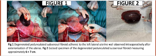
After the secure closure of the uterine incision in standard layers, the fibroid stalk was carefully clamped, excised, and transfixed with delayed absorbable sutures to ensure absolute hemostasis. The excised specimen was subsequently sent for detailed histopathological examination.
Gross pathological examination revealed a well-circumscribed, nodular mass measuring approximately 6 × 7 cm, exhibiting a variable firm-to-soft consistency. On cut section, the mass demonstrated a classic whorled appearance interspersed with extensive areas of dark hemorrhage and cystic degeneration. Microscopic examination of the tissue sections showed interlacing bundles of smooth muscle cells arranged in a characteristic whorled, fascicular pattern, consistent with a leiomyoma. The tumor cells were predominantly spindle-shaped with elongated, cigar-shaped nuclei and abundant eosinophilic cytoplasm. There was no evidence of cellular atypia, increased mitotic activity, or coagulative tumor cell necrosis that would suggest malignancy. Extensive areas of hemorrhagic infarction and hyalinization were noted. Overall, the gross and histopathological findings were definitively consistent with a degenerated uterine leiomyoma.
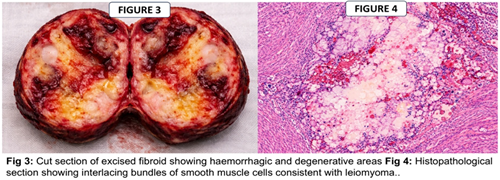
The postoperative period was entirely uneventful. The patient experienced rapid and significant relief from her acute abdominal pain immediately following the surgery. Both the mother and the newborn demonstrated excellent recovery profiles and were discharged in a stable, healthy condition on the fourth postoperative day.
3. Discussion
Uterine fibroids (leiomyomas) are the most common benign tumors of the female reproductive tract and are diagnosed in approximately 1.6% to 10.7% of all pregnancies [2]. While the vast majority of fibroids remain asymptomatic during the gestational period, they may occasionally provoke significant complications depending on their precise size, multiplicity, and anatomical location relative to the placenta and cervix.
Acute pelvic or abdominal pain is the most frequently encountered complication associated with fibroids during pregnancy and is predominantly linked to degenerative changes within the fibroid architecture. Degeneration typically ensues when the fibroid outgrows its vascular supply, a phenomenon exacerbated during pregnancy when the rapid hypertrophy and expansion of the gravid uterus may stretch or compress the feeding vessels. Among the various morphologic subtypes of degeneration, red (carneous) degeneration is the most frequently observed during pregnancy. Pathophysiologically, it results from acute venous stasis and thrombosis, culminating in hemorrhagic infarction within the fibroid. Clinically, this manifests as acute, severe, and unrelenting abdominal pain. This presentation can deceptively mimic other severe causes of an acute abdomen in pregnancy, such as placental abruption, acute appendicitis, adnexal torsion, or the onset of preterm labor, thereby rendering the clinical diagnosis highly challenging [9-11].
In the present case, the patient presented atypically at 37 weeks and 4 days of gestation with severe localized abdominal pain strongly suggestive of a surgical acute abdomen. An additional and notable diagnostic challenge in this scenario was the failure of three prior routine antenatal ultrasonographic examinations to detect the fibroid. Subserosal or pedunculated fibroids can occasionally evade sonographic detection during routine obstetric scans, particularly if they are small in early pregnancy, or if their lateral or fundal positioning makes differentiation from the normal undulating uterine contour or maternal bowel loops difficult.
The patient’s dramatic presentation with persistent, severe abdominal pain at term necessitated prompt surgical intervention. An emergency laparotomy with a lower segment caesarean section was executed due to the acute clinical picture and the advanced gestational age. Intraoperatively, a degenerated pedunculated subserosal fibroid, attached to the uterus by a narrow stalk, was isolated. Pedunculated subserosal fibroids are inherently predisposed to mechanical torsion or vascular avulsion as the uterus rapidly enlarges, which predictably results in ischemic degeneration and the precipitation of severe abdominal pain.
Traditionally, concurrent myomectomy during a caesarean section (caesarean myomectomy) has been strictly discouraged in obstetric practice due to profound concerns regarding intractable intraoperative hemorrhage from the highly vascularized gravid uterus. However, an evolving body of contemporary literature suggests that in carefully selected cases-particularly those involving pedunculated subserosal fibroids-myomectomy can be safely and effectively performed at the time of caesarean section with a minimal risk of hemorrhagic complications. In the case presented, the fibroid was uniquely attached by a narrow pedicle, which facilitated a safe and controlled excision immediately following the closure of the primary uterine incision. Simple clamping, transection, and secure transfixation of the pedicle ensured complete and definitive hemostasis.
This case underscores the critical importance of maintaining a high index of suspicion for degenerated fibroids as a primary differential diagnosis in pregnant patients presenting with an acute abdomen, even in instances where previous comprehensive antenatal imaging has failed to document their presence. Early clinical recognition and the execution of appropriate, decisive surgical management are paramount to mitigating maternal morbidity and ensuring maximally favorable maternal and fetal outcomes.
4. Conclusion
The degeneration of uterine fibroids during pregnancy is an uncommon yet highly significant etiology of acute abdominal pain that can closely mimic other catastrophic obstetric or general surgical emergencies. This case definitively illustrates that fibroids may remain completely undetected during routine antenatal ultrasonography, only to present abruptly with severe, incapacitating pain, particularly in the later stages of pregnancy. Prompt, astute clinical assessment combined with timely surgical intervention is essential to prevent severe maternal and fetal complications. In this instance, an emergency caesarean section followed immediately by the targeted surgical removal of the pedunculated degenerated fibroid resulted in an exceptionally favorable maternal and neonatal outcome. Heightened clinical awareness of this rare presentation can significantly aid obstetricians and emergency physicians in correctly identifying fibroid degeneration as a pivotal differential diagnosis in the evaluation of the pregnant acute abdomen.
Declarations
Ethical Approval
The case report was approved by the Institutional Ethics Committee. Standard protocols for patient confidentiality were strictly maintained.
Consent
Written informed consent was obtained from the patient for the publication of this case report and the accompanying clinical/intraoperative images. A copy of the written consent is available for review by the Editor of this journal.
Conflict of Interest
The author declares that there are no financial or non-financial conflicts of interest regarding the publication of this paper.
Funding
No external funding was received for this study.
Data Availability
All data underlying the results are available as part of the article, and no additional source data are required.